Link Between the Brain and Immune System
Article published by NeuroScience News –
Researchers Find Missing Link Between the Brain and Immune System
Implications profound for neurological diseases from autism to Alzheimer’s to multiple sclerosis.
In a stunning discovery that overturns decades of textbook teaching, researchers at the University of Virginia School of Medicine have determined that the brain is directly connected to the immune system by vessels previously thought not to exist. That such vessels could have escaped detection when the lymphatic system has been so thoroughly mapped throughout the body is surprising on its own, but the true significance of the discovery lies in the effects it could have on the study and treatment of neurological diseases ranging from autism to Alzheimer’s disease to multiple sclerosis.
“Instead of asking, ‘How do we study the immune response of the brain?’ ‘Why do multiple sclerosis patients have the immune attacks?’ now we can approach this mechanistically. Because the brain is like every other tissue connected to the peripheral immune system through meningeal lymphatic vessels,” said Jonathan Kipnis, PhD, professor in the UVA Department of Neuroscience and director of UVA’s Center for Brain Immunology and Glia (BIG). “It changes entirely the way we perceive the neuro-immune interaction. We always perceived it before as something esoteric that can’t be studied. But now we can ask mechanistic questions.”
“We believe that for every neurological disease that has an immune component to it, these vessels may play a major role,” Kipnis said. “Hard to imagine that these vessels would not be involved in a [neurological] disease with an immune component.”
New Discovery in Human Body
Kevin Lee, PhD, chairman of the UVA Department of Neuroscience, described his reaction to the discovery by Kipnis’ lab: “The first time these guys showed me the basic result, I just said one sentence: ‘They’ll have to change the textbooks.’
There has never been a lymphatic system for the central nervous system, and it was very clear from that first singular observation – and they’ve done many studies since then to bolster the finding – that it will fundamentally change the way people look at the central nervous system’s relationship with the immune system.”
Even Kipnis was skeptical initially. “I really did not believe there are structures in the body that we are not aware of. I thought the body was mapped,” he said. “I thought that these discoveries ended somewhere around the middle of the last century. But apparently they have not.”
‘Very Well Hidden’
The discovery was made possible by the work of Antoine Louveau, PhD, a postdoctoral fellow in Kipnis’ lab. The vessels were detected after Louveau developed a method to mount a mouse’s meninges – the membranes covering the brain – on a single slide so that they could be examined as a whole. “It was fairly easy, actually,” he said. “There was one trick: We fixed the meninges within the skullcap, so that the tissue is secured in its physiological condition, and then we dissected it. If we had done it the other way around, it wouldn’t have worked.”
After noticing vessel-like patterns in the distribution of immune cells on his slides, he tested for lymphatic vessels and there they were. The impossible existed. The soft-spoken Louveau recalled the moment: “I called Jony [Kipnis] to the microscope and I said, ‘I think we have something.’”
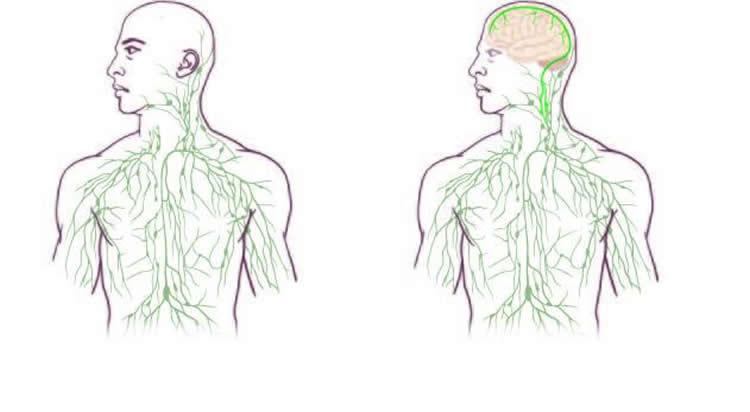
This shows the maps of the lymphatic system: old (left) and updated to reflect UVA’s discovery.
As to how the brain’s lymphatic vessels managed to escape notice all this time, Kipnis described them as “very well hidden” and noted that they follow a major blood vessel down into the sinuses, an area difficult to image. “It’s so close to the blood vessel, you just miss it,” he said. “If you don’t know what you’re after, you just miss it.”
“Live imaging of these vessels was crucial to demonstrate their function, and it would not be possible without collaboration with Tajie Harris,” Kipnis noted. Harris, a PhD, is an assistant professor of neuroscience and a member of the BIG center. Kipnis also saluted the “phenomenal” surgical skills of Igor Smirnov, a research associate in the Kipnis lab whose work was critical to the imaging success of the study.
Alzheimer’s, Autism, MS and Beyond
The unexpected presence of the lymphatic vessels raises a tremendous number of questions that now need answers, both about the workings of the brain and the diseases that plague it. For example, take Alzheimer’s disease. “In Alzheimer’s, there are accumulations of big protein chunks in the brain,” Kipnis said. “We think they may be accumulating in the brain because they’re not being efficiently removed by these vessels.” He noted that the vessels look different with age, so the role they play in aging is another avenue to explore. And there’s an enormous array of other neurological diseases, from autism to multiple sclerosis, that must be reconsidered in light of the presence of something science insisted did not exist.
About this neuroscience research
The findings have been published online by the prestigious journal Nature and will appear in a forthcoming print edition. The article was authored by Louveau, Smirnov, Timothy J. Keyes, Jacob D. Eccles, Sherin J. Rouhani, J. David Peske, Noel C. Derecki, David Castle, James W. Mandell, Lee, Harris and Kipnis.
Funding: The study was funded by National Institutes of Health grants R01AG034113 and R01NS061973. Louveau was a fellow of Fondation pour la Recherche Medicale.
Source: Debra Kain – University of Virginia Health System
Image Source: The image is credited to the University of Virginia Health System
Original Research: Abstract for “Structural and functional features of central nervous system lymphatic vessels” by Antoine Louveau, Igor Smirnov, Timothy J. Keyes, Jacob D. Eccles, Sherin J. Rouhani, J. David Peske, Noel C. Derecki, David Castle, James W. Mandell, Kevin S. Lee, Tajie H. Harris and Jonathan Kipnis in Nature. Published online June 1 2015 nature14432
Abstract
Structural and functional features of central nervous system lymphatic vessels
One of the characteristics of the central nervous system is the lack of a classical lymphatic drainage system. Although it is now accepted that the central nervous system undergoes constant immune surveillance that takes place within the meningeal compartment1, 2, 3, the mechanisms governing the entrance and exit of immune cells from the central nervous system remain poorly understood4, 5, 6. In searching for T-cell gateways into and out of the meninges, we discovered functional lymphatic vessels lining the dural sinuses. These structures express all of the molecular hallmarks of lymphatic endothelial cells, are able to carry both fluid and immune cells from the cerebrospinal fluid, and are connected to the deep cervical lymph nodes. The unique location of these vessels may have impeded their discovery to date, thereby contributing to the long-held concept of the absence of lymphatic vasculature in the central nervous system. The discovery of the central nervous system lymphatic system may call for a reassessment of basic assumptions in neuroimmunology and sheds new light on the aetiology of neuroinflammatory and neurodegenerative diseases associated with immune system dysfunction.
“Structural and functional features of central nervous system lymphatic vessels” by Antoine Louveau, Igor Smirnov, Timothy J. Keyes, Jacob D. Eccles, Sherin J. Rouhani, J. David Peske, Noel C. Derecki, David Castle, James W. Mandell, Kevin S. Lee, Tajie H. Harris and Jonathan Kipnis in Nature. Published online June 1 2015 nature14432
Feel free to share this Neuroscience News.
![[RLLY] Lymplex](/web/image/product.template/3257/image_1920/%5BRLLY%5D%20Lymplex?unique=ae82545)
![[RLXP] Xeneplex](/web/image/product.template/3508/image_1920/%5BRLXP%5D%20Xeneplex?unique=ae82545)
![[RLZO] Zoiben](/web/image/product.template/3517/image_1920/%5BRLZO%5D%20Zoiben?unique=ae82545)