Gunther Enderlein
Gunther Enderlein (1872-1968) saw the healthy host as filled with primitive life forms which he called – Colloids of Life or Protits. These reside in the red cells, white cells, in plasma and all other body fluids and tissues, are 0.01 micron in radius (about the size of a virus), and the larger forms of these can be seen under any microscope’s high power, oil immersion lens, as tiny dots, rolling, always moving. They are seen best with a dark-field microscope as tiny shining, moving points. They are visible because they move. For more information about Gunther Enderlein see the Explore Magazine.
Gunther Enderlein was the last of the old pleomorphists and has done the most exhaustive and comprehensive compilation and study of this information to date. The basis for Enderlein’s work was the book by the French researcher A. Bchamp, titled Mycrozymas. Enderlein devoted the bulk of his scientific work which stretched for more that 40 years, to the complex question of pleomorphism, symbiosis and cyclogeny (the cycles organisms go through) of microorganisms.
From this he published his chief work, BAKTERIEN-CYCLOGENIE (publisher W. de Gruyter & Co. Berlin, 1925). In it he presented arguments and proofs for pleomorphism, which have to this date remained undefeated. This book has just become available in English from Enderlein Enterprises, Inc., P.O. Box 704, Mt. Vernon, WA 98273, phone 360-424-6029. That there has been very little about any of this available in English has been part of the problem for sure.
The following information and pictures, except where otherwise indicated, are from the book Blood Examination in Darkfield according to Prof. Dr. Gunther Enderlein, available from Semmelweis-B Verlag, D-27316 Hoya, Germany, compiled by Dr. med. Maria-M Bleker, 1993. This book is also available, in English and German, at the above address. In the following, unidentified quotes are placed for material taken from this book. Maria Bleker gives seminars in Darkfield Microscopic examination and has been one of the chief people responsible for introducing Pleomorphism to America. This has not been an easy job.
“According to Enderlein, ‘germs’ are not representing unchanging organisms that are independent of each other, but altogether they form a singular, common cycle, which has its origin in the colloidal, albuminoid substances called Protits that are contained inside of each particular cell.”
Enderlein called these Protits, ENDOBIONTS (from the Greek endo- internal and bios- life). We can never separate ourselves from them. We coexist in a mutually symbiotic (means we live together, helping each other) relationship. We give them a vehicle for life, they give us blood forms like platelets, without which we couldn’t exist (platelets are formed from the Protits, not in the bone marrow as taught by modern science). The endobiont appears in all mammalian species and has shown evidence through some of its developmental forms to be of a plant nature. Our symbiotic union with them evidently occurred millions of years ago as our species grew into existence. Without some blood clotting mechanism in place, mammals could never have evolved.
When the host is in health, Protits live in symbiotic relationship with the tissue cells, and maintain the health of and regenerate all organs, we live together and help each other. They are the smallest unit of life, not the cell.
They are physical life per se.
In the blood and tissue of humans and animals, there live microbes (Protits) which are normally harmless and which maintain diverse regulatory mechanisms. According to Prof. Enderlein, these ‘Endobionts’ are usually nonpathogenic phases of the mold fungus Mucor racemosus Fresen, Aspergillus niger van Tieghem or Penicillium notatum. One does not see these fungal forms in the blood of a living being, these are just what they look like when cultured in a laboratory or what the Protits devolve to in a corpse. The mummies in Egypt are composed totally of Protits. Put the dust from these mummies in water and there they are, under the microscope, turning, moving, the Protits.
The Law of Cyclogeny
As with microbes the above Protits are subject to the Law of Cyclogeny, which means, their development cycles through the following stages: primitive phase (Protit) to bacterium to fungus, whereby the virulence and pathogenicity of the parts of the cycle increases with the rising developmental phase.
This means, they develop from the apathogenic (can’t make you sick), non-motile, tiniest albuminoid, particle (Protit) – which is to be classified in size with the viruses (0.01 m) – via the nonvirulent chondrit stage into the following parasitic, pathogenic (can make you sick) stages.
As the patient becomes ill and therefore acid, microorganisms form, in quantum-biological leaps, depending on the pH of the nutritive medium or the internal milieu. The microorganisms get more dangerous as one goes from the strongly alkaline high of the pH-value towards the ever decreasing pH-value of the acid side.
This happens in the following manner. AS THE INTERNAL MILIEU BECOMES MORE ACID, the Protits first begin to join into threads (tails or Filum) that sprout globules, primitive granules (heads). These, Enderlein, called SYMPROTITS.
As from Dr. Med. Maria-M Bleker’s, Blood Examination in Darkfield according to Prof. Dr. Gunther Enderlein;
“In the new formation of filum and head, atomic-physical and quantum-biological factors play a decisive role. This is visible from the sudden occurrences by the leap of these new formations. The formational processes of a filum with a head resulting in what is called the CHONDRIT, occurs within the smallest fraction of a second, which is therefore not observable by the eye looking through the microscope; the new developmental forms are suddenly there. The Protits come together and join in the following manner:
Protits can NATIONALIZE or come together in three ways. They can form:
- A one dimensional arrangement. This results in a shorter or longer thread, the FILUM; its diameter is that of the protit, namely 0.01 mm. However, it can constantly increase in thickness, after its formation.
- A two dimensional arrangement of the protits, into fine, skin-like surfaces that are found, for example, in the spermit (bacteriophage) as swarmer-heads (see below). This is one filum with one Protit head. The SYMPROTIT is simply a larger head than the Protit or the Spermit head.
- A three-dimensional arrangement, namely into more or less tiny granules, the physiologic, often spherical SYMPROTITS which become the primary nuclei of the bacterial cell they are turning into.”
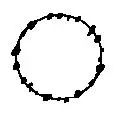
Formation of Filum
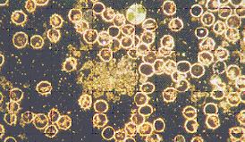
The formation of these Fila threads can be observed only in the dark field, sometimes this occurs very rapidly, right before your eyes, in extremely ill patients.
The fila in this picture are the thread like structures that fill the background. These are what cause blood to clot. The circles are red blood cell. The circles that are stacked together in the left hand picture are red blood cells that have stuck together.
As from Maria Bleker’s book;
“It is the first of the primitive phases that occurs through the change in fila according to the formula x:2x. Because the x shows very great differences in the lengths, the filit-phase comprises very large numbers of individual phases. These threads begin to appear soon after the blood is drawn. While watching under the microscope the threads just pop into view, increasing in length in quantum type jumps, 2X long then 4X, then 8X, 16X, etc.. Actually, there appears to be constant quantum fluctuations between these formed products. Thereupon follow the primitive stage FILIT, the ongoing change between the FILUM and a filum-piece of double its length.”
If a person is totally healthy, none of these Fila will form. If there is no appearance of Fila after two days, that person is totally healthy. Personally, I have never seen this, perhaps in a new born baby.
Base Powder reverses this process.
Actually, we had a patient one time in whom the Fila started forming immediately. This is very unusual as usually it takes some hours for this process to start. This patient was very ill with extremely acid blood and cancer and we never saw him again. How fast the Fila form is a very good indicator of how ill you are as it is a direct reflection of how acid you are.
Formation of Symprotits
Next, the Filit makes a quantum jump and unites with or produces, instantaneously sprouts, a SYMPROTIT HEAD. The above tail simply develops a head, in a quantum fashion and poof….there is a filum thread with a symprotit head on it – out of ‘nowhere’…?
This is called the CHONDRIT (see below), a filum with a symprotit head. You have sperm, or do you? The similitude among sexual processes in Nature is pervasive and awesome. The snake that ate itself. In fact sometimes these threads or fila begin to move like a flagella and you have what is called SPERMIT. This is pleomorphism and acid base is the key.
“The spermits of the microbes are tiny swarmers that consist of a tiny symprotit heads and filum flagellie, which enables them to copulate with all pathological symprotits and all the bacterial and fungal forms within the same cycle. The consequence of such copulation of bacterial and fungal-nucleic apparatus is naturally that the bacteria and fungi immediately become dissolved and they degrade back into more spermits and protits/chondrits. The presence of spermits is a good thing as these forms attack anything (higher pathogenic forms). Spermits cannot be photographed because of their minute size and intense mobility. They represent nothing but a readiness to meet an alarming situation. ”
Enderlein first discovered this process in 1916 with his work on typhus. He rediscovered in the blood of these patients, with darkfield, “the tiniest moving beings”, this having been seen before by Bchamp and others. Enderlein saw too that these tiny moving beings “entered into union with higher organized bacteria”. When this happened the bacteria that had joined with the tiny beings “became instantly invisible. Enderlein surmised sexual processes, through which came about, not higher forms (as in embryonal development) but lower forms that were invisible to the eye in the light microscope. These vigorously moving elements had flagella.” As indicated above he named these Spermits.
Symprotits
Symprotit phase- acid base imbalance.
This shows the variation in size of the Protits. The small dots in this picture are Protits, the larger dots are the Symprotits. The red circles are red blood cells and the mass in the middle of the slide are disintegrating blood cell. The heads can only be seen here, not the tails.
Symprotits are conglomerated Protit heads that form varying sized globules or heads. As the Protits group together the heads get larger and larger. As stated, the tails really can’t be seen in these pictures. The Symprotits then that we see under the microscope are enlarged Protit type forms occurring in various sizes. They are always moving too, compared to other things seen under the microscope that look similar and don’t move.
The small dots in this picture are larger and of different diameters than the ones in the above picture. The Protits have stuck together and formed the heads of the next larger “nationalization” or grouping of the Protits. Each advancement to a higher stage or VALENCY as it is called represents an increasing degree of pathogenicity. They become more dangerous.
Then, these conglomerated Protits (Symprotits) use more protein colloid, Protits, for their advancement, depositing it first in large numbers right on its surface as nutritional reserves. This reserved living, protein colloid grows even larger, surrounding the symprotit sphere with more and more substance. These become Macrosymprotits (macro – large, symprotits). The acid base imbalance is becoming worse.
Macrosymprotits
These represent exceptionally large spheres of purely nucleic protein. They can be found free, or connected with the filum, or in the elements of tissues and cells of the host. In connection with the filum, the fila are, then, usually very mobile.
“One can tell by the diverse sizes of ‘free’ symprotits that are present, whether one is dealing with normal forms or abnormal ones. Namely, in that case, the formula is also x:2x, which means the presence of identical spheres plus other ones of twice their size. When several sphere-phases are found side-by-side, they will be indicated by a larger number of varieties in size, which is the more common condition. They just jump from one size to the other, no period in-between that is observable. The resulting forms are the large, heads or primitive granules called Macrosymprotits.”
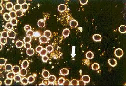
Macrosymprotit
The large dot the arrow is pointing to is a Macrosymprotit. Notice the difference in size of the “tiny dots” in this picture.
As the internal milieu becomes acid, as the pH decreases, the primary tiny lumps and their tails, the Protits begin to increase in size by sticking together in a three dimensional arrangement; first one, then two, then eight, then sixteen, then thirty-two according to the formula, x:2x, where x is the number of Protits coming together. These increase in quantal jumps too. You can watch it under the microscope; first there is 1 Protit, then 2, then 4, 8… and so on, all lumped together.
Free Condrit Phase
The Chondrit Phase begins with the above enlargement of the Symprotits and of their Fila. The Symprotits and their tails join end to end giving the Chondrit forms a beaded appearance. This gives them a lively mobility as they have an even denser arrangement of tiny symprotits along the length of the fila.
“The primitive stage CHONDRIT can be seen most frequently constantly changing between FILUM and PRIMITIVE GRANULE (Symprotit). Depending on the size of this tiny primitive granule (between 0.02 m and 1m), very diverse valences or degrees of pathogenicity may occur at this stage.”
FREE CHONDRIT PHASE
Free Chondrit Phase
The large dots in this picture are Macrosymprotits. The beaded threads like the one the lower arrow is pointing to are free Chondrit heads on Fila threads. Some have coalesced into, thick rid shaped forms, (see below). The large circles are red blood cells.
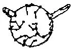
The Chondrit Stage
RBC with Chondrit growing out of it
This is the Chondrit stage growing out from a red blood cell edge. It has many Symprotit heads on a File thread. This stage is very strong and mobile. This thread will close end to end and form a circle. This will become a bacterial cell as below.
The lower valenced Chondrits are not pathogenic and do not make you sick. In fact, these low valenced Chondrits plus the Protit discussed above and the Protitit (the forms that are so small that they can’t be seen with the light microscope). The Protit and Protitit have even lower valency or pathogenicity than the Chondrits and these three are what are used as Isopathic medicine. These Chondrits, being quite mobile, seek out higher valenced microorganisms such as bacteria and fungal forms. The Chondrit forms copulate and fuse genetic material with these more pathogenic organisms and they then break all apart into Protits as described.
The Chondrit Stage begins with that sphere of the developmental growth of the endobiont, in which only the low-valenced phases have full apathogenicity and all higher phases reach pathogenicity to an ever rising degree. The higher valenced forms of these Chondrits have more Symprotit heads, they are bigger and more varied in size among other things. The Free Chondrits are considered to be the viral phase and in their higher valenced forms they are pathogenic as such.
“THE ONLY EXCEPTIONS ARE THE VERY FIRST PRIMITIVE STAGES which are the PROTIT and the CHONDRITS that are of lowest valences. They are entirely nonvirulent and the play a REGULATORY role toward the higher and more pathogenic stages by decomposing these through copulatory processes. In that sense, these stages are termed REGULATORS.”
Isn’t this all rather amazing? “Sperms”, doing what they do and breaking down higher and more dangerous forms of its conglomerated self, back into itself. The snake eating its tail for sure. This is Isopathic medicine.
The Birth of a Cell (bacterial spheres)
Next, if the internal milieu, Pishinger’s Space, becomes worse, more acid, polluted, the beaded, Free Chondrits, form circles (closed loops) by hooking end to end and looping around to complete a circle. These circles are composed of symprotit heads, distributed around the ring made of fila.
Symprotit heads distributed around a ring made of fila.
These symprotit heads will fuse together and form the nucleus of the developing bacterial cell.
Then, these small symprotit heads move together and fuse forming the nucleus (called the Mych)

Mych
and you have the newborn cell!
Bacterial Spheres
The medium sized circles like the arrow is pointing, looking like bubbles, are Bacterial Spheres (Mychits) or spherical, primary bacterial cells. The large circles are red blood cells and the mass in the middle is a disintegrating white blood cell with the many ring forms being extruded from it.
The above are the growth forms of the primitive phases (the Protit, Symprotit, Macrosymprotit and lower valenced Chondrits). These come together and form the cell.
“Each higher developmental step represents the nationalization or coming together of these growth forms. In this way, the symprotit uses the protit, namely a protein-colloid, for its advancement, depositing it at first in large numbers right on its surface as nutritional reserves. This reserved living, protein colloid grows ever larger, surrounding the symprotit sphere more and more. By this process, the first cell has come to be, which is a spherical primary cell, the bacterial cell, the MYCHIT. By this process, the symprotit became the primary nucleus, the MYCH, and from the reserve material collection of living colloids came the CELL PLASMA of the primary cell, the Mychit.”
“Reserve substances, which form a more or less thick layer, may be deposited around the primary nucleus. These primary nuclei with their covering of reserve substances completely correspond physiologically to the fatty substances in higher organisms. To the very largest percentage of cases, these reserve materials consist of LIPOIDS and also NUCLEIC ACID DERIVATIVES.”
Development of Bacterial Rods
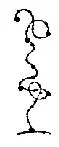
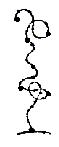
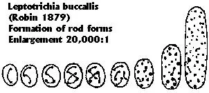
Through division, the above Bacterial Sphere becomes the source of a micrococcus with two nuclei (DIPROTIT). From them, bacteria with 4 – 8 nuclei develop, and finally a bacillus with 16 and more nuclei. On the other hand, the nucleus divides and the ring can elongate and turn into rod forms. This is a picture drawn while looking though a microscope in 1879. The bacterium, Leptotrichia buccalis, the usually harmless bacteria found in our mouths, is seen here to go from the round coccal form into a rod form. In the process the nucleus divides and becomes many.
Formation of Bacterial Rods
To summarize, the small nuclei or tiny symprotits of the Chondrit around the edges of the ring in these pictures move together and make one nucleus. The ring can enlarge and become a coccus or round, ball shaped germ or it can elongate and turn into a bacterial rod. The Symprotit gathers Protits to it to feed it and to form the cellular plasma. The nucleus gets larger as the tiny symprotits coalesce and the ring gets larger as the cell plasma fills the cell up.
Coccus type germs are the streptococcus, gonococcus, staphlococcus and the like. Rod type germs are E. coli, pseudomonis, and so on. These forms actually come out of the cells themselves, red or white in the blood. The blood, when it starts to break down first begins a fermentation process that can lead to blood clots or rigor mortuus, two sides of the same process.
Two bacterial rods exuding from RBC
Red blood cell from a patient with stomach cancer. There are two bacterial rods exuding from it. The nuclei are stacked one on top of the other.
The following pictures show rod forms budding off of or extruding from red blood cells. You see round buds pinching off of blood cells. You see long hollow tubes that have come out of cells and long, beaded strings of Chondrit forms that can be the pathogenic viral forms. There are also some short stubby rods that are typical of the ones we see in medical, micro biology labs. Acid base imbalance is severe.
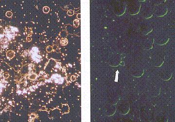
Budding Cells
The multitude of various size dots in the left hand picture are Symprotits and Macrosymprotits, Symplasts (heads and tails hooked end to end, not yet pulled together into bacterial forms) and Free Chondrits (the beaded, long thread like forms). The right hand picture shows buds of bacterial forms coming out of red blood cells.
What makes these life forms, Protits, ‘advance’ so rapidly? (I prefer to use the word degenerate or involve, devolve – rather than advance or evolve.)
“Our civilization causes or facilitates this through artificial fertilizers, preservatives, coloring substances, air pollution, etc., but in the very first place stands our false nutrition, which literally “fattens” the Endobiont by its high-content in protein and sugar. Animal protein fattens the Protit. As soon as the balance in the blood serum between mineral salts (bases, alkali) and acids has become disturbed toward the acidic side through long-continued, antibiological nutrition, the above things start sticking together. The endobiont literally feeds off animal protein and gets bigger.”
“The endobiont is the ROBBER OF PROTEIN. The only non-plant protein that can be taken in larger amounts, is the protein of the milk, and that in its acid form, such as cottage cheese and other forms of cheese. Farmers Cheese is the best available in supermarkets. These lactic proteins have developed a special accomplishment in the course of endless time, namely the capacity for producing a specific protein synthesis, which does not give the endobiont an opportunity to feed on.”
The normal healthy Protit is made up of vegetable protein!
“The hydrogen-ion concentration (pH) of the blood gets shifted through the Endobiont, whereby it must be especially emphasized that the Endobiont expressly devours protein. It is understandable that these facts create ever enlarging conditions of the endlessly ongoing development of the Endobiont.”
The Anartatic Law of Enderlein
The dependency of the development of microorganisms upon the pH (acid base) of the nutritive medium or the internal milieu is a FUNDAMENTAL LAW called the Anartatic Fundamental Law by Enderlein. As from Blutuntersuchung Im Dunkelfeld nach Prof. Dr. Gunther Enderlein;
The Anartatic Fundamental Principle:
“For the nationalization (coming together of) of comparative-morphologic units into higher and highest developmental phases, the specific acids PRODUCED by each individual microorganism are the CAUSAL reason for the changes of the internal milieu in the pH, and that is tending to the ACIDIC side. In other words: the RISING steps of the total cyclogeny are accompanied by and dependent on the PROPORTIONATELY DESCENDING pH. That is it demonstrates the summary of the ASCENDING developmental tendency with the ever more DESCENDING pH-value.”
An interesting point here is that one can never force an advancement by increasing the acidity of the culture medium. This won’t, in and of itself, cause the Protits to stick together and acquire tails and form spheres with nuclei on their edges that finally turn into bacterial and then fungal forms.
“On the other hand, if one alkalinizes a culture medium already containing fully developed bacterial or fungal forms by adding a little bacterial material or parts of fungal mycelia to a hanging drop of 5% sodium carbonate, which is a strongly alkaline medium with a high pH value, one can immediately observe the formation of the primitive stages, namely in the CHONDRIT STAGE.”
So higher forms are readily ‘decomposed’ back into the Chondrit Stage, by making them alkaline. So too, if one withdraws blood from a vein through a tube and then run the blood directly into a sugar-water solution with 40% ethyl alcohol in it, without exposing it to air, as per Louis Pasteur and Antoine Bechamp, it will begin to ferment. This fermentation produces the acids it needs to continue its evolution, de-evolution, involution, namely lactic and citric acids. The red blood cells begin to decompose. All the above things happen and what you end up with is nothing but Protits. It takes a month or so. Very interesting experiment.